Muscles of the head and neck worksheet – Embark on an in-depth exploration of the muscles of the head and neck with this comprehensive worksheet. Delve into the intricate network of muscles that orchestrate facial expressions, mastication, and neck movements, gaining a profound understanding of their anatomical relationships, functions, and clinical significance.
Prepare to unravel the complexities of the craniofacial musculature, deciphering the actions, innervation, and blood supply of each muscle. Engage in a journey of anatomical discovery, mastering the fundamentals of head and neck anatomy.
Introduction to the Muscles of the Head and Neck: Muscles Of The Head And Neck Worksheet
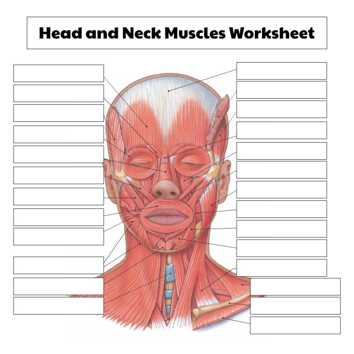
The muscles of the head and neck are responsible for a wide range of essential functions, including facial expressions, chewing, swallowing, and head and neck movements. These muscles are innervated by various cranial and cervical nerves and are supplied with blood by branches of the external carotid artery.
Muscles of the Scalp
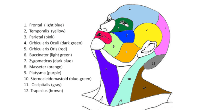
The muscles of the scalp are responsible for scalp movement and are innervated by branches of the facial nerve. The main muscles of the scalp include the:
- Frontalis muscle: Originates from the galea aponeurotica and inserts into the skin of the forehead. It elevates the eyebrows and wrinkles the forehead.
- Orbicularis oculi muscle: Surrounds the eye and inserts into the skin around the orbit. It closes the eyelids.
- Corrugator supercilii muscle: Originates from the medial aspect of the orbit and inserts into the skin of the eyebrow. It draws the eyebrows together.
Muscles of the Face
| Muscle | Origin | Insertion | Action | Innervation |
|---|---|---|---|---|
| Orbicularis oris muscle | Mandibular and maxillary bones | Skin around the mouth | Closes the lips | Facial nerve |
| Buccinator muscle | Mandible and maxilla | Skin and mucous membrane of the cheek | Compresses the cheek and helps in chewing | Facial nerve |
| Zygomaticus major muscle | Zygomatic bone | Skin of the corner of the mouth | Elevates the corner of the mouth and produces a smile | Facial nerve |
Muscles of Mastication
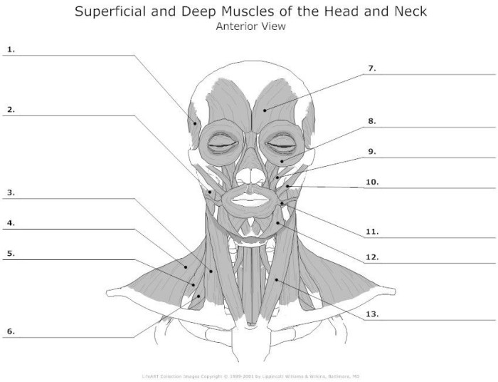
The muscles of mastication are responsible for chewing and are innervated by the mandibular nerve, a branch of the trigeminal nerve. The main muscles of mastication include the:
- Masseter muscle: Originates from the zygomatic arch and inserts into the mandible. It elevates the mandible.
- Temporalis muscle: Originates from the temporal fossa and inserts into the coronoid process of the mandible. It elevates and retracts the mandible.
- Medial pterygoid muscle: Originates from the medial surface of the lateral pterygoid plate and inserts into the medial surface of the mandible. It elevates the mandible and helps in protrusion.
- Lateral pterygoid muscle: Originates from the lateral surface of the lateral pterygoid plate and inserts into the articular disc and neck of the mandible. It helps in protrusion and side-to-side movement of the mandible.
Muscles of the Neck
- Sternocleidomastoid muscle: Originates from the sternum and clavicle and inserts into the mastoid process of the temporal bone. It flexes and rotates the neck.
- Trapezius muscle: Originates from the occipital bone, cervical vertebrae, and thoracic vertebrae and inserts into the clavicle, acromion process, and spine of the scapula. It elevates and retracts the scapula and extends the head.
- Scalene muscles: Originate from the transverse processes of the cervical vertebrae and insert into the ribs. They flex and rotate the neck.
FAQ Overview
What is the function of the temporalis muscle?
The temporalis muscle elevates the mandible, contributing to jaw closure and clenching.
How many muscles are responsible for facial expressions?
There are 26 muscles involved in facial expressions, each contributing to a unique range of movements.
