Cells of this tissue shorten to exert force, a fundamental mechanism underlying a wide range of biological processes. This process, mediated by the cytoskeleton, particularly actin and myosin, enables cells to generate contractile forces that drive essential functions such as muscle contraction, cell migration, and tissue morphogenesis.
Cells have evolved specialized adaptations to enhance their force-generating capacity, including structural modifications, cell-cell interactions, and extracellular matrix interactions. The regulation of cell shortening involves intricate molecular pathways and signaling mechanisms, ensuring precise control over force generation in response to various stimuli.
Understanding Cell Shortening and Force Generation

Cell shortening is a fundamental cellular process that enables cells to exert force and perform various functions. It involves the coordinated action of cytoskeletal components, primarily actin and myosin, to generate contractile forces.
Actin filaments form the backbone of the cytoskeleton and provide structural support to the cell. Myosin motors, attached to actin filaments, use ATP hydrolysis to slide along them, causing the filaments to shorten. This shortening generates force that can be transmitted to the extracellular environment or to other cellular structures.
Examples of cell types that exhibit cell shortening for force generation include muscle cells, which contract to generate movement, and smooth muscle cells, which regulate blood flow and other physiological processes.
Cellular Adaptations for Force Generation, Cells of this tissue shorten to exert force
Cells have evolved structural adaptations to enhance their ability to shorten and generate force effectively. These adaptations include:
- Cell shape:Cells with elongated or spindle-shaped morphologies can generate more force than spherical cells due to the increased overlap of actin and myosin filaments.
- Cell-cell interactions:Adhesion molecules, such as integrins, connect cells to each other and to the extracellular matrix, providing a stable substrate for force generation.
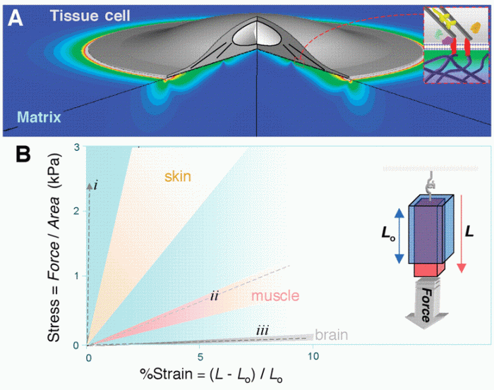
- Extracellular matrix:The extracellular matrix, composed of proteins and carbohydrates, provides mechanical support and resistance to cell shortening, allowing cells to generate more force.
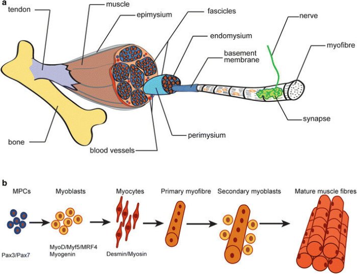
Specialized cell types have evolved unique adaptations for force generation. For example, muscle cells have highly organized sarcomeres, the contractile units that generate muscle force, while smooth muscle cells have a network of actin and myosin filaments that allows them to maintain sustained contractions.
Regulation of Cell Shortening
Cell shortening is tightly regulated by a complex network of molecular pathways and signaling mechanisms. These include:
- Calcium ions:Calcium influx into the cell triggers the activation of myosin motors, initiating cell shortening.
- Kinases:Protein kinases, such as Rho kinase, phosphorylate and activate myosin motors, enhancing their contractile force.
- Other regulatory proteins:Calmodulin, tropomyosin, and cofilin are among the many proteins that modulate the activity of actin and myosin, fine-tuning cell shortening.
Cells respond to various stimuli by adjusting the activity of these regulatory pathways, thereby modulating their force-generating capacity. For example, muscle cells increase calcium influx upon nerve stimulation, leading to muscle contraction, while smooth muscle cells respond to hormonal signals by altering the activity of myosin motors.
Applications of Cell Shortening in Tissue Function
Cell shortening plays a crucial role in various tissue functions, including:
- Muscle contraction:The coordinated shortening of muscle cells generates force for movement, locomotion, and other physiological processes.
- Cell migration:Cells shorten to move through the extracellular matrix during processes such as wound healing and immune responses.
- Wound healing:The shortening of myofibroblasts, specialized cells in wound tissue, generates tension that helps close wounds.
Tissue-specific adaptations in cell shortening contribute to overall tissue function. For example, the highly organized sarcomeres in muscle tissue allow for rapid and powerful contractions, while the slower, sustained contractions of smooth muscle cells enable the regulation of blood flow and other physiological processes.
Manipulating cell shortening has potential therapeutic applications in tissue repair and regeneration. For instance, enhancing cell shortening in damaged muscle tissue could improve muscle function, while inhibiting cell shortening in pathological conditions, such as fibrosis, could prevent tissue damage.
Question & Answer Hub: Cells Of This Tissue Shorten To Exert Force
What is the role of actin and myosin in cell shortening?
Actin and myosin are key cytoskeletal proteins that interact to generate contractile forces. Actin filaments slide past myosin filaments, causing cell shortening and force generation.
How do cells regulate cell shortening?
Cell shortening is regulated by various molecular pathways and signaling mechanisms, including calcium ions, kinases, and other regulatory proteins, which control the activity of cytoskeletal components.
What are the applications of cell shortening in tissue function?
Cell shortening plays a crucial role in muscle contraction, cell migration, wound healing, and other tissue functions. Tissue-specific adaptations in cell shortening contribute to overall tissue function.


